Venous Ulcers Cause: Venous hypertension and valve failure poor venous return Typical features: Location: medial gaiter area (ankle/lower leg), rarely toes Appearance: shallow, irregular edges Exudate: often heavy Skin changes: oedema, hyperpigmentation, lipodermatosclerosis Pain: mild to moderate, improves with elevation Management focus: Compression therapy (once arterial disease excluded) Skin care and oedema control Wound dressings to manage exudate Arterial Ulcers Cause: Peripheral arterial disease (reduced blood flow) Typical features: Location: toes, forefoot, heel, pressure points Appearance: punched-out, deep, often necrotic Exudate: minimal Skin changes: cool, pale, shiny, hair loss Pain: severe, worse at night or when leg is elevated Management focus: Urgent vascular assessment Revascularisation where possible Avoid compression until perfusion is confirmed Why this distinction is critical Compression heals venous ulcers but can destroy an ischaemic limb Debridement may be appropriate in venous ulcers but dangerous in arterial disease without blood flow Dressing choice, escalation, and prognosis differ significantly Key message Treating an ulcer without understanding its cause is treating blind
What is more, mechanistic study indicated that p53 activation was involved in the inhibitory effect of SLC7A11 induced by FA administration, which could be suppressed by LA supplementation
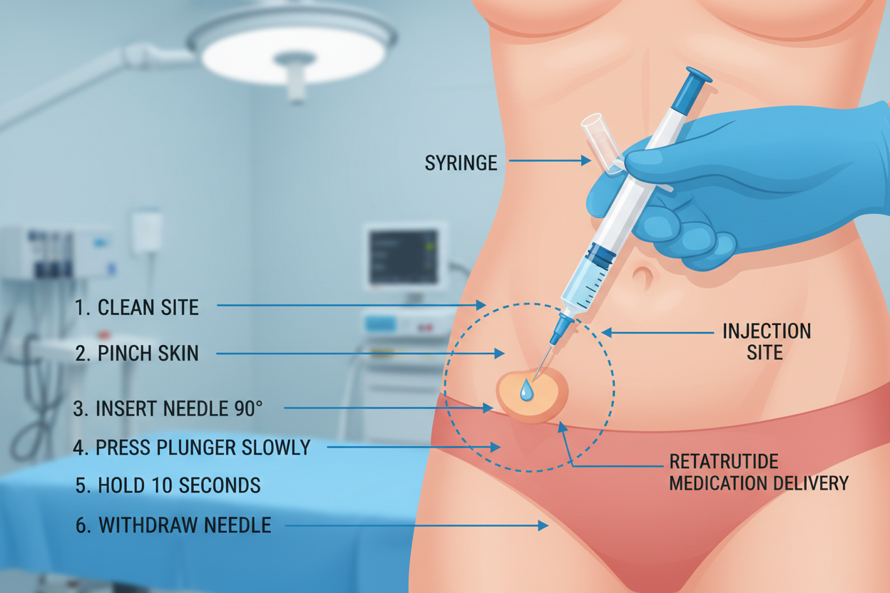
Patients are provided with a detailed explanation of the treatment process , including what to expect at each stage
Just dont
doi: 10.1165/rcmb.2005-0336RC
10.1002/14651858.CD009384.pub3 290